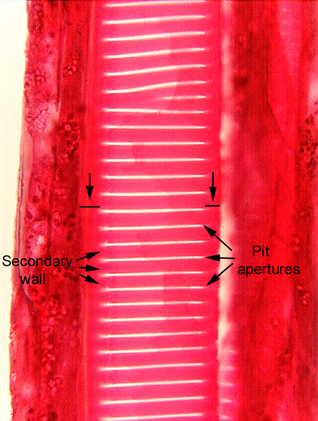
 Fig.
7.2-4.
Longitudinal section of fern stem (Pteridium). This micrograph shows
excellent scalariform
pitting. Each long, horizontal white area is the pit
aperture (actually, we are looking through the two apertures of each
pit-pair) and the red is the Safranin-stained secondary wall. Note that almost
all of the primary wall is covered with secondary wall in a cell like this –
there is so much secondary wall that the cell is strong enough to provide
considerable support to the stem. The two lines pointed out by the arrows
indicate the corners of the cell that are not penetrated by the scalariform
pits. Because these corners are not pitted, they are especially strong and
prevent the cell from being stretched by the surrounding tissues.
Fig.
7.2-4.
Longitudinal section of fern stem (Pteridium). This micrograph shows
excellent scalariform
pitting. Each long, horizontal white area is the pit
aperture (actually, we are looking through the two apertures of each
pit-pair) and the red is the Safranin-stained secondary wall. Note that almost
all of the primary wall is covered with secondary wall in a cell like this –
there is so much secondary wall that the cell is strong enough to provide
considerable support to the stem. The two lines pointed out by the arrows
indicate the corners of the cell that are not penetrated by the scalariform
pits. Because these corners are not pitted, they are especially strong and
prevent the cell from being stretched by the surrounding tissues.