Up
Primary xylem
Oak wood
Leaf vein
Vein ends
Bean seed
Pine tracheids, xs
Fern TE, xs
Fern, TE, mag
Annular walls
Annular, stretched
Annular, narrow
Scalariform walls
Scalar., narrow
CBP, pine
CBP, dicot
CBP, irregular
Contact faces
Pits, side view
CBP, pine, xs
CBP,angio, xs
CBP, fern, xs
Contact face, xs
Simple perf. plate 1
Simple perf. plate 2
Pitted perf. plate
Perf. plate & helix
Perf. plate, face
Perf. plate, mag
Perf. plate, section
Perf. plate rim
Perf. plate & wall
Scalariform Per plate
Primary xylem
Vessel sizes
Fern TE
Pine needle
VE precursor, ls
Protoxylem
9 Contact faces
VE precursor, xs
Precursor 2
Torn vessel
Torn vessel 2
| |
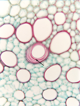 Fig.
7.3-4. Transverse section of vascular bundle in parsnip (Pastinaca).
This micrograph shows a simple perforation plate lying parallel to the plane of
sectioning, but it is in a vessel element with a helical secondary wall rather
than a pitted one as in Fig. 7.3-3. The
arrow marks the portion of wall that is the perforation plate, the other red
bands are helices of secondary wall. Fig.
7.3-4. Transverse section of vascular bundle in parsnip (Pastinaca).
This micrograph shows a simple perforation plate lying parallel to the plane of
sectioning, but it is in a vessel element with a helical secondary wall rather
than a pitted one as in Fig. 7.3-3. The
arrow marks the portion of wall that is the perforation plate, the other red
bands are helices of secondary wall.
Most
of the tissue is slightly out of focus, as is even part of the perforation
plate. The perforation lies deep within the section, so by focusing on it, all
the rest of the tissue is out of focus. The fact that part of the perforation is
in focus and part is not indicates that it is tilted, not perfectly parallel to
the plane of sectioning.
|