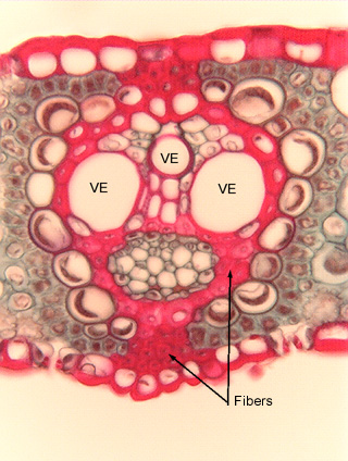
 Fig.
7.1-3. Transverse section of leaf of sugar cane (Saccharum
officinale). Students often study the xylem of leaf
veins (vascular bundles), and this vein in a sugar cane leaf is
rather representative of monocots. It is common to encounter a sheath of fibers,
which are easy to confuse with tracheary elements because they have thick walls
that are usually stained red. But most tracheary elements are much wider than
fibers, as are two of the three vessels here. The narrowest vessel is just a
little wider than a fiber, and it is often very difficult to be certain if some
cells are fibers or tracheary elements.
Fig.
7.1-3. Transverse section of leaf of sugar cane (Saccharum
officinale). Students often study the xylem of leaf
veins (vascular bundles), and this vein in a sugar cane leaf is
rather representative of monocots. It is common to encounter a sheath of fibers,
which are easy to confuse with tracheary elements because they have thick walls
that are usually stained red. But most tracheary elements are much wider than
fibers, as are two of the three vessels here. The narrowest vessel is just a
little wider than a fiber, and it is often very difficult to be certain if some
cells are fibers or tracheary elements.
The set of brownish cells below the two large vessels is the phloem (larger, empty-looking cells are sieve tube members, the small cells with contents are companion cells).