Up
Primary xylem
Oak wood
Leaf vein
Vein ends
Bean seed
Pine tracheids, xs
Fern TE, xs
Fern, TE, mag
Annular walls
Annular, stretched
Annular, narrow
Scalariform walls
Scalar., narrow
CBP, pine
CBP, dicot
CBP, irregular
Contact faces
Pits, side view
CBP, pine, xs
CBP,angio, xs
CBP, fern, xs
Contact face, xs
Simple perf. plate 1
Simple perf. plate 2
Pitted perf. plate
Perf. plate & helix
Perf. plate, face
Perf. plate, mag
Perf. plate, section
Perf. plate rim
Perf. plate & wall
Scalariform Per plate
Primary xylem
Vessel sizes
Fern TE
Pine needle
VE precursor, ls
Protoxylem
9 Contact faces
VE precursor, xs
Precursor 2
Torn vessel
Torn vessel 2
| |
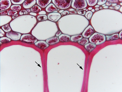 Fig.
7.1-7b.
Magnification of vessel elements in fern (Pteridium). The three large
cells at the bottom of the micrograph are vessel elements. The two arrows
indicate the contact faces (it is possible to see that the “wall” between
two cells is actually two walls and a middle lamella). Although this looks like
an ordinary wall that you might see between two fibers, the paleness in each
indicates that there is a scalariform opening there. The opening happens to be a
perforation in this Pteridium, not a pit. Fig.
7.1-7b.
Magnification of vessel elements in fern (Pteridium). The three large
cells at the bottom of the micrograph are vessel elements. The two arrows
indicate the contact faces (it is possible to see that the “wall” between
two cells is actually two walls and a middle lamella). Although this looks like
an ordinary wall that you might see between two fibers, the paleness in each
indicates that there is a scalariform opening there. The opening happens to be a
perforation in this Pteridium, not a pit.
|