 Fig.
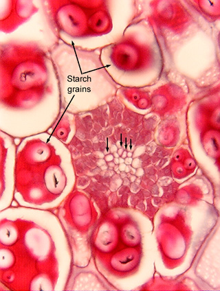
7.1-5. Transverse section of vascular bundle in a
bean seed (Phaseolus vulgaris). This section passes through one of the
cotyledons of a bean (the cotyledons are the two large halves of beans, peas,
and peanuts for example). Four arrows indicate four tracheary elements; notice
that each is narrower -- much narrower -- than a single starch grain.
Considering that starch grains are subcellular organelles, each of these
tracheary elements is narrower than a small part of an ordinary parenchyma cell.
Fig.
7.1-5. Transverse section of vascular bundle in a
bean seed (Phaseolus vulgaris). This section passes through one of the
cotyledons of a bean (the cotyledons are the two large halves of beans, peas,
and peanuts for example). Four arrows indicate four tracheary elements; notice
that each is narrower -- much narrower -- than a single starch grain.
Considering that starch grains are subcellular organelles, each of these
tracheary elements is narrower than a small part of an ordinary parenchyma cell.
Are these tracheary elements tracheids or vessels? Can't tell? Neither can I. With a longitudinal section and an excellent high power lens, we might just be able to see perforations if they are present and oriented properly. But even a tiny fragment of starch could hide the miniscule perforations these might have. And even if we could see a perforation, it might appear to be nothing more than a blemish, an artifact. We might look right at a perforation and still not be certain if these are vessel elements or tracheids.