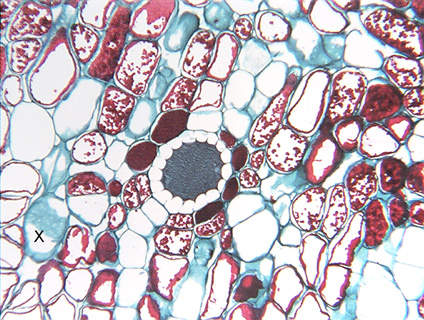
 Fig.
3.2-8.
Transverse section of root of Clusia (a tree from tropical Central
America with no common name in English). The secretory
product of this duct was preserved by the fixation process and has
taken up a blue stain (secretory products are often dissolved during processing,
so ducts typically appear empty on microscope slides). This duct is lined by one
layer of secretory parenchyma cells that appear to be completely empty. Many of
the surrounding cells have deposits of tannins that have stained as red
particles or as solid red masses. In some, the tannins occur only along the
wall, so at first glance the tannin might appear to be a red-stained secondary
wall, but a secondary wall would not be as rough or irregular as these tannin
deposits. The cell at xxx has a bit
of front (or back) wall present, showing the primary pit fields as small whitish
dots and splotches.
Fig.
3.2-8.
Transverse section of root of Clusia (a tree from tropical Central
America with no common name in English). The secretory
product of this duct was preserved by the fixation process and has
taken up a blue stain (secretory products are often dissolved during processing,
so ducts typically appear empty on microscope slides). This duct is lined by one
layer of secretory parenchyma cells that appear to be completely empty. Many of
the surrounding cells have deposits of tannins that have stained as red
particles or as solid red masses. In some, the tannins occur only along the
wall, so at first glance the tannin might appear to be a red-stained secondary
wall, but a secondary wall would not be as rough or irregular as these tannin
deposits. The cell at xxx has a bit
of front (or back) wall present, showing the primary pit fields as small whitish
dots and splotches.