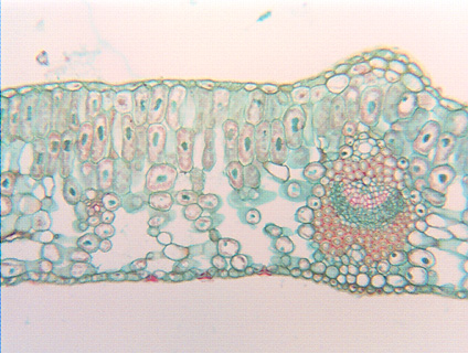
 Fig.
3.3-2. Transverse section of leaf of ivy (Hedera). Like leaves
of most species, this ivy leaf consists of almost pure parenchyma, except for
its veins. Even though ivy leaves are somewhat tough and leathery, their upper
and lower epidermis and all the photosynthetic tissues are composed of
parenchyma cells. We can consider the parenchyma to be structural
because it makes up most of the bulk of the leaf, but the photosynthetic cells
are simultaneously synthetic parenchyma, and the epidermis cells are boundary
parenchyma. Notice that the lower half of the leaf has much larger intercellular
spaces – is more aerenchymatic – than the upper half. This slide is unusual
in that nuclei are stained green rather than the more typical red, although both
colors are artificial – unstained nuclei are colorless.
Fig.
3.3-2. Transverse section of leaf of ivy (Hedera). Like leaves
of most species, this ivy leaf consists of almost pure parenchyma, except for
its veins. Even though ivy leaves are somewhat tough and leathery, their upper
and lower epidermis and all the photosynthetic tissues are composed of
parenchyma cells. We can consider the parenchyma to be structural
because it makes up most of the bulk of the leaf, but the photosynthetic cells
are simultaneously synthetic parenchyma, and the epidermis cells are boundary
parenchyma. Notice that the lower half of the leaf has much larger intercellular
spaces – is more aerenchymatic – than the upper half. This slide is unusual
in that nuclei are stained green rather than the more typical red, although both
colors are artificial – unstained nuclei are colorless.