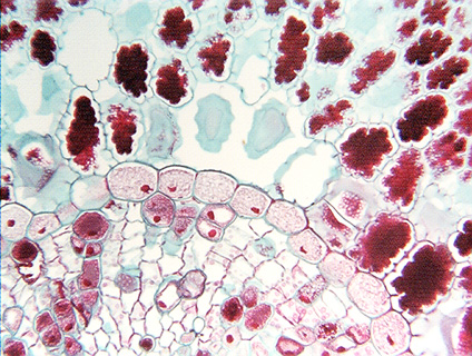
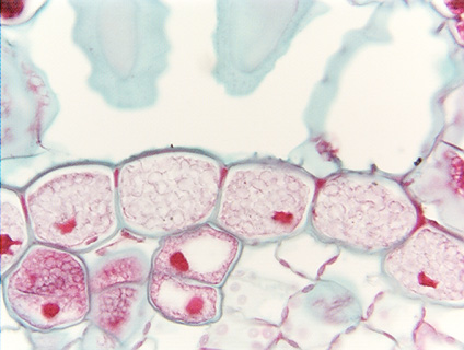
Fig. 3.4-2a and b. Transverse section of a needle leaf of pine (Pinus). The long, cylindrical needle leaves of pine have an outer chlorenchyma made up of lobed cells (upper part of low magnification view) and a central vascular bundle (lower part of low magnification view). The two tissues are separated by a boundary parenchyma known as an endodermis. The endodermis is a single layer of cells (high magnification view) that fit against each other tightly with no intercellular spaces, and which have a waterproof Casparian strip that runs completely around each cell. Endodermis would be useless as a boundary if it had intercellular spaces.
Even though these endodermis cells have an abundance of starch grains, red-stained nuclei are visible in several. The nuclei are lumpy and irregular, not spherical – this is due to being deformed by having starch grains pressing against them.