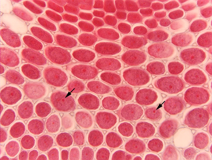
 Fig. 3.1-8. Transverse
section of the funiculus (the stalk that attaches a seed to a fruit as it
develops) in bean (Phaseolus vulgaris). These parenchyma cells have
contents that have been stained red, making the intercellular
spaces quite visible. The cytoplasmic staining is so intense it is
difficult to see nuclei, but in many cells the very dark red, small dot is a
nucleolus (arrows), and the nucleus can be detected in some of the cells as a mass with
slightly different color surrounding the nucleolus.
Fig. 3.1-8. Transverse
section of the funiculus (the stalk that attaches a seed to a fruit as it
develops) in bean (Phaseolus vulgaris). These parenchyma cells have
contents that have been stained red, making the intercellular
spaces quite visible. The cytoplasmic staining is so intense it is
difficult to see nuclei, but in many cells the very dark red, small dot is a
nucleolus (arrows), and the nucleus can be detected in some of the cells as a mass with
slightly different color surrounding the nucleolus.