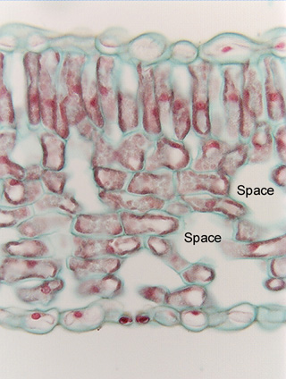
 Fig. 3.2-1.
Transverse section of leaf of common barberry (Berberis vulgaris).
All cells in this micrograph are parenchyma cells. The topmost and bottommost
layers are the upper and lower epidermises of the leaf, and all cells in between
are technically known as mesophyll (Chapter 12: Leaf). These mesophyll cells
have very obvious contents, most of which have been stained red, which is very
common in slides of plant tissue (the stain Safranin O stains many different
organelles, turning them all red). Cells of the lower epidermis appear rather
empty, which makes it easy to identify the nucleus in four cells -- the nuclei
are round and are a uniform red. The cells just below the upper epidermis are
the palisade parenchyma: each cell is columnar and most have a large red-stained
nucleus near the middle of the cell. Almost all of the rest of the red-stained
material in those cells consists of chloroplasts. The chloroplasts were green
when the leaf was alive, but the alcohol used to preserve the leaf tissues
dissolves chlorophyll, bleaching the chloroplasts which then become stained red.
Between the palisade parenchyma cells and the lower epidermis are spongy
mesophyll cells. Their chloroplasts are so abundant they make it difficult to
see the nuclei; on a microscope, the nuclei can be identified by focusing up and
down with the fine focus. The primary walls have been stained blue-green, and
all are very thin. Intercellular spaces are large in spongy mesophyll but
smaller in palisade parenchyma (some of the large white areas in the palisade
are regions where the cells were cut away by the microtome knife, and although
they appear large, they are very thin).
Fig. 3.2-1.
Transverse section of leaf of common barberry (Berberis vulgaris).
All cells in this micrograph are parenchyma cells. The topmost and bottommost
layers are the upper and lower epidermises of the leaf, and all cells in between
are technically known as mesophyll (Chapter 12: Leaf). These mesophyll cells
have very obvious contents, most of which have been stained red, which is very
common in slides of plant tissue (the stain Safranin O stains many different
organelles, turning them all red). Cells of the lower epidermis appear rather
empty, which makes it easy to identify the nucleus in four cells -- the nuclei
are round and are a uniform red. The cells just below the upper epidermis are
the palisade parenchyma: each cell is columnar and most have a large red-stained
nucleus near the middle of the cell. Almost all of the rest of the red-stained
material in those cells consists of chloroplasts. The chloroplasts were green
when the leaf was alive, but the alcohol used to preserve the leaf tissues
dissolves chlorophyll, bleaching the chloroplasts which then become stained red.
Between the palisade parenchyma cells and the lower epidermis are spongy
mesophyll cells. Their chloroplasts are so abundant they make it difficult to
see the nuclei; on a microscope, the nuclei can be identified by focusing up and
down with the fine focus. The primary walls have been stained blue-green, and
all are very thin. Intercellular spaces are large in spongy mesophyll but
smaller in palisade parenchyma (some of the large white areas in the palisade
are regions where the cells were cut away by the microtome knife, and although
they appear large, they are very thin).