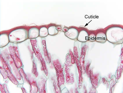
 Fig.
3.4-1. Transverse section of leaf of Peucephyllum (no common
name). The layer of large round cells is the epidermis, a type of boundary
parenchyma. The thick, red-stained layer is the cuticle, composed of
the water-proof material cutin. In this species, as in most, epidermis cells
secrete cutin mostly to their outer wall, not the inner one. There is some
deposition on the outer parts of the radial walls also. Because these epidermis
cells have thin primary walls, they are parenchyma cells. The columnar cells in
the lower half of the micrograph are palisade parenchyma cells, filled with
red-stained chloroplasts. The two small cells with red contents in the center of
the epidermis are the two guard cells on either side of a stomatal pore. Other
than the stomatal pore, there are no intercellular spaces between epidermis
cells.
Fig.
3.4-1. Transverse section of leaf of Peucephyllum (no common
name). The layer of large round cells is the epidermis, a type of boundary
parenchyma. The thick, red-stained layer is the cuticle, composed of
the water-proof material cutin. In this species, as in most, epidermis cells
secrete cutin mostly to their outer wall, not the inner one. There is some
deposition on the outer parts of the radial walls also. Because these epidermis
cells have thin primary walls, they are parenchyma cells. The columnar cells in
the lower half of the micrograph are palisade parenchyma cells, filled with
red-stained chloroplasts. The two small cells with red contents in the center of
the epidermis are the two guard cells on either side of a stomatal pore. Other
than the stomatal pore, there are no intercellular spaces between epidermis
cells.