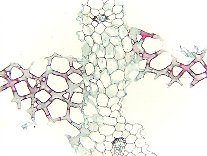
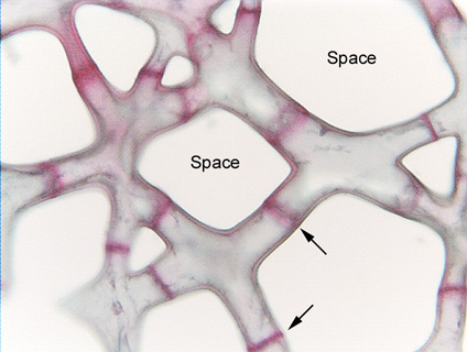
Fig. 3.3-6. Transverse section of leaf of cattail (Typha). Leaves of cattail have a very light-weight construction that is easy to see by just cutting or tearing open a leaf and looking at it closely. The giant air spaces are separated by plates that are not merely thin (one cell thick), even the plates themselves are mostly air. They are composed of branched parenchyma cells like the ones shown here. The red bands (two are indicated by arrows) are cross walls separating one cell from another, and each cell has from four to six “arms.” These parenchyma cells developed from small cells that had a cubical shape when the leaf was just a tiny primordium. But as the leaf grew, neighboring cells became partially unglued from each other in certain spots, and those spots developed into intercellular space. In the spots where the cells remained glued together by their middle lamella, those areas were stretched into arms. Try to imagine exactly which walls were adjacent to which when the tissue was younger.