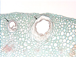
Fig. 9.3-7. Transverse section of flower petal of lemon (Citrus limon). Probably everyone is familiar with the large oil pockets in the peels of oranges and lemons and other citrus fruits. The petals of citrus flowers also have oil glands, although they are smaller than those of the peels, and fit into a micrograph more easily. The low magnification view shows two glands , one appearing to have more contents than the other. It may be that the section is near the front or back of the cavity: unlike the ducts in pine and parsnip, these are chambers – almost perfectly spherical, so many sections are glancing sections at or near the back or front of the chamber. If the section contained all of the front or back region, the gland would appear to be solid.
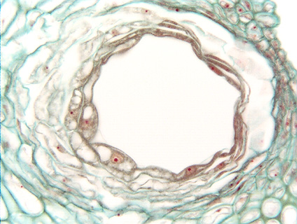
The high magnification shows a thick epithelium, with the innermost cells being densely cytoplasmic and with prominent nuclei. Outside of the innermost layer are many other layers of epithelial cells. These glands grow by recruitment: a small set of cells begins producing oil, and they stimulate their neighbors to begin doing the same thing. As the neighbor cells become active, they in turn activate the ones just to the exterior of themselves. Once mature, oil cells break down, creating the lumen; this is holocrine secretion. The gland grows larger as more petal cells become converted to oil-producing cells. This recruitment occurs symmetrically, so the gland grows as a spherical chamber; if it occurred on just one or two sides, it would grow as a tube. See Figs. 9.7 and 9.23 in Plant Anatomy (Mauseth).