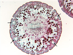
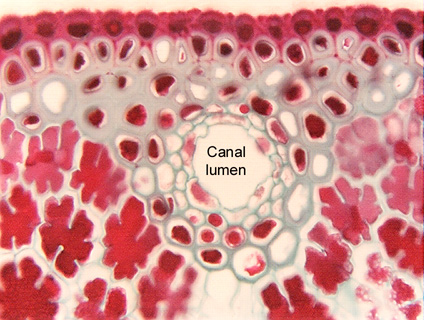
Fig. 9.3-6. Transverse section of needle leaf of pine (Pinus). The needles of pines and many other conifers have resin canals with complex epithelia. There are several layers of epithelium cells, the innermost layer consists of parenchyma cells with prominent nuclei, the outer layers are cells with thick walls. The low magnification view shows that the canals are located near the surface of the leaf, and at fairly close intervals around the perimeter (remember how slender pine needles are) – it would be almost impossible for even a small animal to bite into this needle without encountering the bitter resins.
Pine needles have a variety of interesting features. One is the epidermis made up of cells with thick walls, another is the presence of lobed parenchyma cells making up much of the leaf (all the red-stained cells).